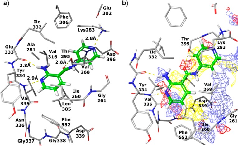
Figure 1.
Compound 2 (in green) modeled into X-ray structure of PKC-ι. (a) Hydrogen bonds between 2 and protein residues are shown as yellow dashed lines; (b) a grid is superimposed on the binding site showing areas favorable for binding ligand atoms with the following properties: hydrophobic (yellow), acceptor or negatively charged (red), and donor or positively charged (blue).

