Fig. 2.
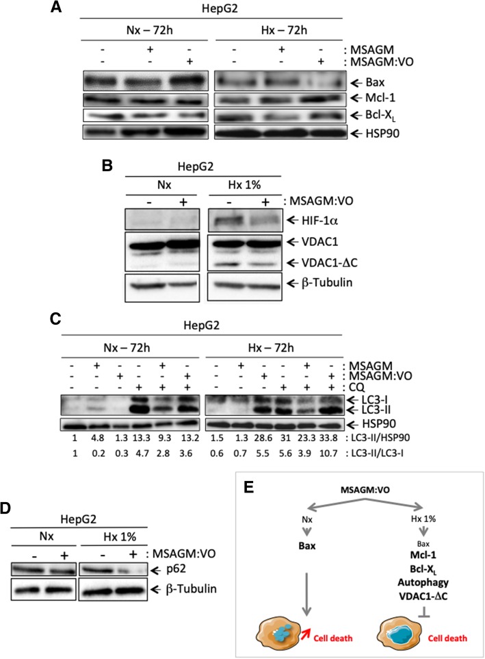
Mechanisms activated in hypoxia protect cells from the apoptosis triggered by MSAGM:VO. a and b HepG2 were incubated in Nx or Hx 1% for 72 h in the absence (−) or presence (+) of MSAGM or MSAGM:VO. Cell lysates were analyzed by immunoblotting for (a) Bax, Mcl-1 and Bcl-XL and (b) HIF-1α and VDAC1. HSP90 and β-tubulin were used as a loading control. c HepG2 cells were incubated in Nx or Hx (1% O2) for 72 h in the absence (−) or presence (+) of MSAGM or MSAGM:VO and in the absence (−) or presence (+) of chloroquine (CQ). Cell lysates were analyzed via immunoblotting for LC3-I and -II. HSP90 was used as a loading control. d HepG2 cells were incubated in Nx or Hx 1% for 72 h in the absence (−) or presence (+) of MSAGM:VO. Cell lysates were analyzed via immunoblotting for p62. β-tubulin was used as a loading control. e Schematic representation of a pathway for hypoxic protection of HepG2 cells in the presence of MSAGM:VO

