
Figure 6.
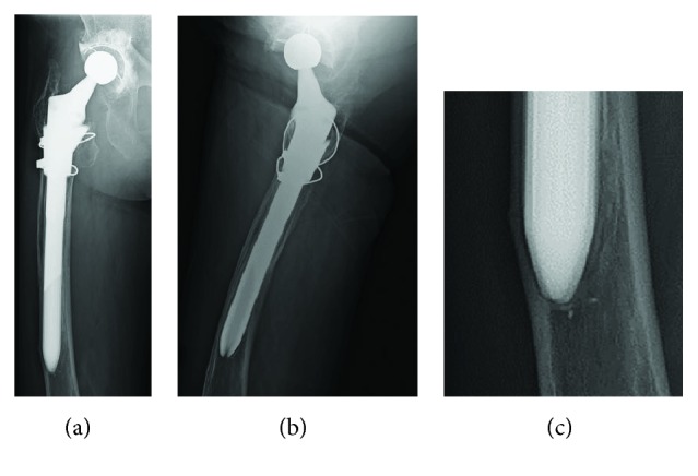
(a) Anteroposterior radiograph, (b) mediolateral radiograph, and (c) magnified anteroposterior radiographs of the fracture line. Five months before hospitalization, the cortical bone was thinning slightly in proximity to the tip of the stable femoral stem and the transverse lucency was admitted on the outside.