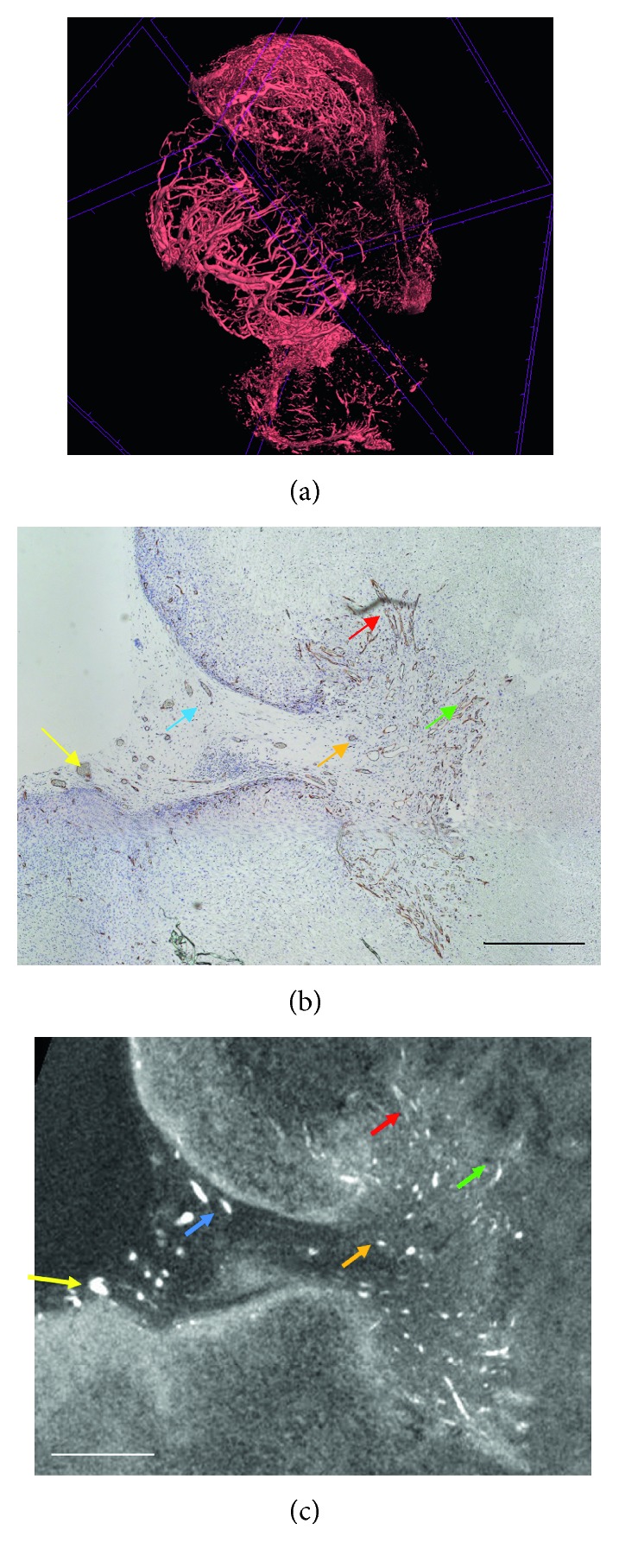
Figure 2.

CE-CT images of vascularization in a tumour xenograft sample, adapted from Kerckhofs et al. [59]. (a) 3D rendering of the vasculature in a tumour xenograft, stained with Hf-WD POM; 3D scale bar = 100 µm. (b) The CD31 stained section. (c) The corresponding CE-CT cross section through the tumour xenograft. The brown colour in the histological section indicates CD31-positive blood vessels. The white colour in the CE-CT image represents red blood cells in the blood vessels. The coloured arrows show corresponding blood vessels in both images. Scale bars = 100 µm.
