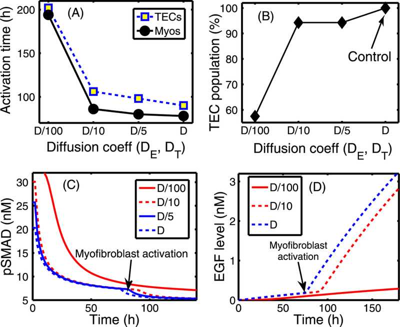
Fig. 19.

The effect of changing the diffusion coefficients of EGF (DE) and TGF-β (DT). (A) Activation time of TECs and myofibroblasts as a function of diffusion coefficients (De, DT) of regulating molecules (EGF and TGF-β). The diffusion coefficients of both EGF and TGF-β were reduced by 5-, 10-, and 100-fold compared to the control case. There are delays in activation of TECs and myofibroblasts when the diffusion coefficients are reduced. (B) The TEC population at day 10 compared to the control (arrow). (C) The time course of pSmad concentration at one EC near (0.4, 0.5) for the four cases of decreased diffusion coefficients considered. Myofibroblasts are activated (arrow) around 194, 86, 80, 78 h, respectively, when TGF-β reaches the threshold, and the pSmad level decreases slowly for smaller values of the diffusion coefficients, leading to a delay of the TEC activation time. For instance TEC is activated around 106 h in the case of 10-fold reduction relative to 90 h in the control case. (D) Initially slow increase of EGF concentration at (0.42, 0.5) near breast duct membrane begins to accelerate (arrow) when myofibroblasts are activated at t = 78, 80, 86 h for relatively smaller reductions of the diffusion coefficients (D, D/5 ,D/10), respectively. However, myofibroblasts are activated at a much later time (194 h) for the significantly stiffened tissue (very small diffusion, D/100)
