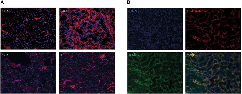
FIGURE 1.
Tomato-positive fluorescent signal in healthy and injured kidneys in renal fibrosis models. (A) Fluorescent tomato signal of FoxD1(-derived) positive cells in FoxD1-Cre;tdTomato in the UUO model and the IRI model. CLK, contralateral (healthy) kidney. (B) Ten days after UUO, co-staining of tomato Red with α-SMA indicates the majority of myofibroblasts are derived from FoxD1-positive cells.

