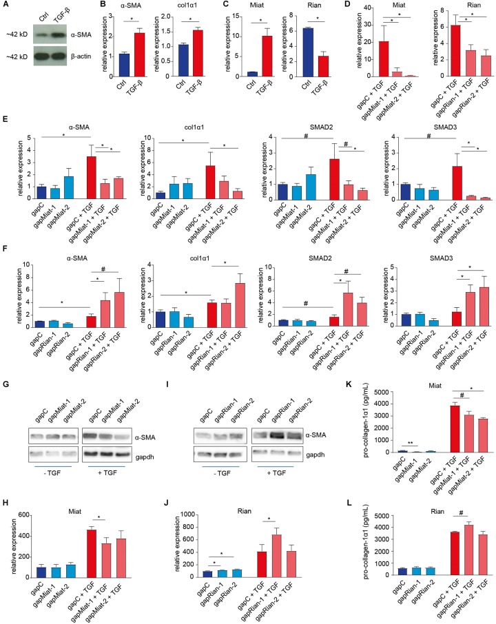
FIGURE 4.
Involvement of lncRNAs Rian and Miat in myofibroblast formation. (A,B) Mouse fibroblasts (NIH3T3 cells) were used to model TGF-β induced myofibroblast formation, illustrated by the induction of α-SMA expression, determined by western blot (A) and α-SMA and collagen-1α1 gene expression (B), determined by qRT-PCR. (C) Similar to the in vivo situation, Rian expression is decreased, while Miat expression in increased, upon TGF-β treatment of mouse fibroblasts. (D) Knockdown of Miat and Rian using gapmers upon treatment of 3T3 cells with TGF-β, as determined by qRT-PCR. N = 3 (three independent experiments) for panels A–D. (E) Knockdown of Miat using gapmers resulted in a decrease of α-SMA, col1a1, SMAD2, and SMAD3 gene expression upon treatment of 3T3 cells with TGF-β, as determined by qRT-PCR. (F) Knockdown of Rian using gapmers increased α-SMA, col1a1, SMAD2, and SMAD3 gene expression upon treatment of 3T3 cells with TGF-β, as determined by qRT-PCR. (G,H) Representative western blot images (G) and quantification (H) indicate that Miat knockdown results in decreased α-SMA levels. (I,J) Representative western blot images (I) and quantification (J) indicate that Rian knockdown results in increased α-SMA levels in non-stimulated cells, while only gapmer 1 increases α-SMA in the presence of TGF-β. N = 6 (six independent experiments) for panels E–J. (K,L) Pro-collagen-1α1 ELISA indicated Miat knockdown decreases pro-collagen levels (K), while gapmer 1 for Rian increases pro-collagen-1α1 levels in the presence of TGF-β (L). N = 3 (three independent experiments) for panels K,L. #P < 0.01, ∗P < 0.05, ∗∗P < 0.01. Data are represented as mean ± SEM.

