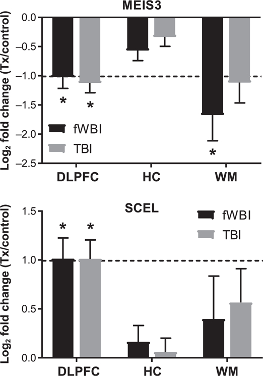
FIG. 3.

MEIS3 and SCEL are differentially expressed within the dorsolateral prefrontal cortex of both fWBI and TBI animals. MEIS3 was downregulated within the dorsolateral prefrontal cortex (log2 fold change: –1.02) and white matter (log2 fold change: –1.67) of animals receiving fWBI, and the dorsolateral prefrontal cortex (log2 fold change: –1.12) of animals receiving TBI. SCEL was upregulated within the dorsolateral prefrontal cortex in animals receiving fWBI (log2 fold change: 1.01) and animals receiving TBI (log2 fold change: 1.01). Bars denote mean log2 fold change and error bars are set to log2 fold standard error. Dashed line indicates log2 fold change of ±1.0. DLPFC = dorsolateral prefrontal cortex; HC = hippocampus; WM = temporal white matter. *FDR adjusted P value ≤ 0.05
