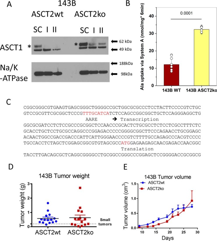
Figure 4.
Compensation of ASCT2 ablation. A, ASCT1 surface protein expression was analyzed by Western blotting in 143B cells (ASCT2wt and ASCT2ko). To identify the ASCT1-specific band (asterisk), RNAi using a scrambled control RNA (SC) and two ASCT1-specific constructs (I and II) was used. Na+/K+-ATPase was used as the protein-loading control. B, induction of alanine uptake mediated by system A (SNAT1/2) in ASCT2ko cells was analyzed by measuring alanine uptake in the presence or absence of 10 mm MeAIB. The net MeAIB-sensitive uptake +/− S.D. is shown (p = 0.0001, n = 8). C, promoter fragment and exon 1 of ASCT1. The AARE and the start codon (ATG) are highlighted in red. D and E, xenografts of ASCT2wt and ASCT2ko 143B cells were established in nude mice, and growth was monitored for 28 days. Tumor weights were measured upon euthanasia (D). Tumor volumes were measured by calipers every 2–3 days. Raw data are shown for tumor weights; mean ± S.E. is reported for tumor volume (E).

