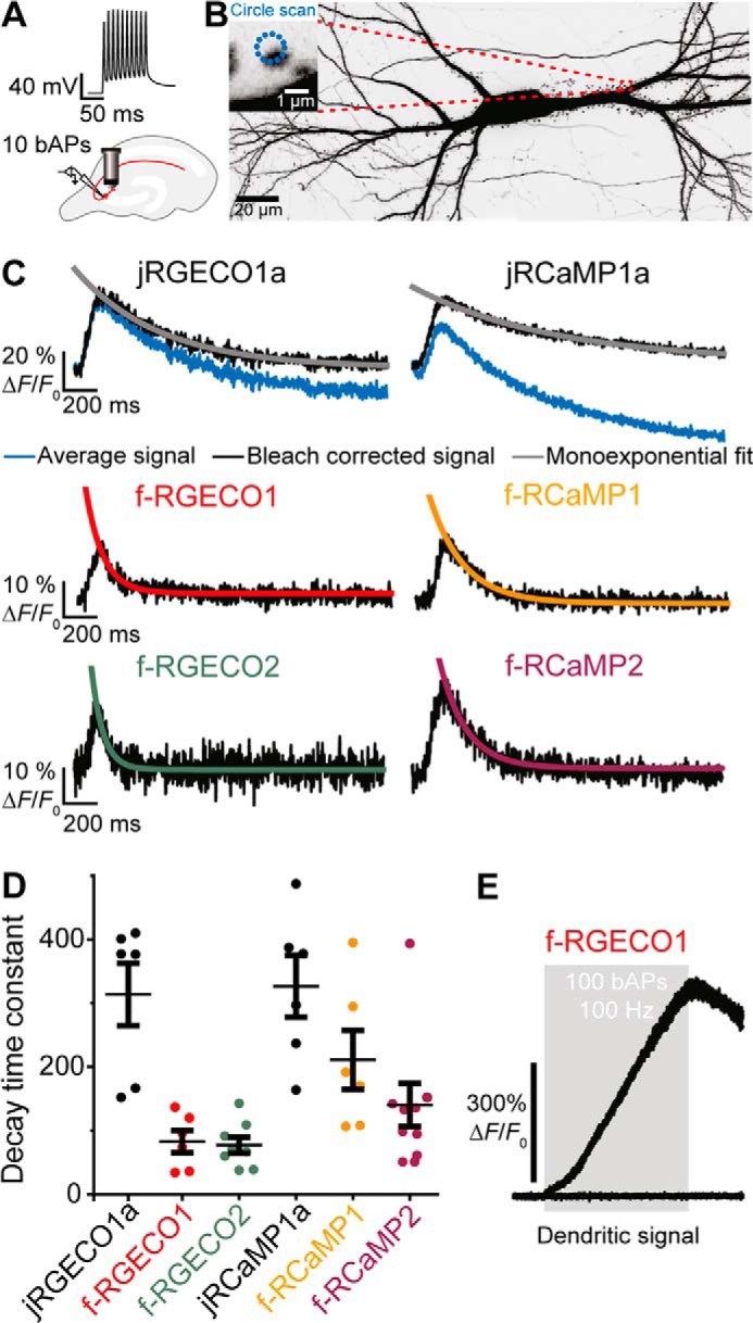
Figure 3.

Spine Ca2+ transients in response to backpropagating action potentials. A, backpropagating action potentials are elicited in a transfected CA3 pyramidal cell by somatic current injections. Ca2+ transients are simultaneously optically recorded from a single spine with two-photon imaging. B, maximum intensity projection of two-photon image stacks of CA3 pyramidal neuron expressing jRGECO1a and GFP, 8 days after electroporation. Green fluorescence intensity is shown as inverted gray values. The scale bar represents 20 μm (neuron) and 1 μm (single spine). C, decay time measurements after bleaching correction (red trace) for individual experiments (five trials averaged per spine) by single exponential fit. D, decay time constants measured in individual spines in response to 10 backpropagating action potentials of CA3 neurons expressing jRGECO1a (τoff = 314 ± 49 ms; n = 6 spines); f-RGECO1 (τoff = 83 ± 17 ms; n = 6 spines); f-RGECO2 (τoff = 77 ± 13 ms; n = 8 spines); jRCaMP1a (τoff = 327 ± 49 ms; n = 6 spines); f-RCaMP1 (τoff = 211 ± 46 ms; n = 6 spines); and f-RCaMP2 (τoff = 140 ± 34 ms; n = 9 spines). The values are expressed as means ± S.E. E, dendritic Ca2+ transients in response to 100 backpropagating action potentials at 100 Hz (train duration, 1 s; gray box) measured in the apical dendrite of a CA3 neuron expressing f-RGECO1. The low affinity of f-RGECO1 prevents the signal from reaching a saturating plateau even in response to a train of 100 backpropagating action potentials.
