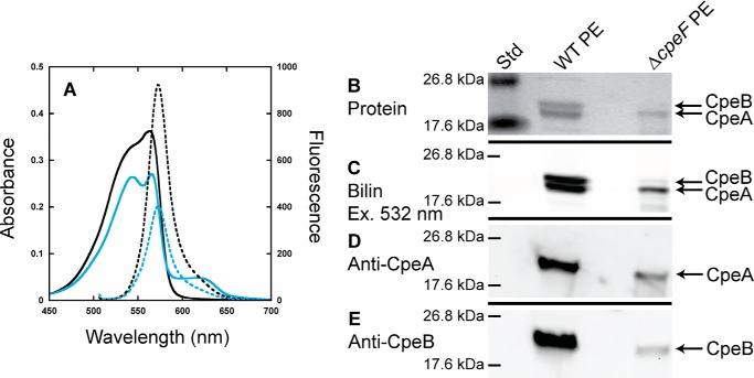
Figure 2.
Analyses of PE purified from WT and ΔcpeF. A, absorbance (solid lines) and fluorescence emission (dashed lines; excitation set at 490 nm) spectra of PE purified from WT (black lines) and ΔcpeF (cyan lines) F. diplosiphon cells grown in green light. B, the Coomassie-stained SDS-polyacrylamide gel for PE purified from WT and ΔcpeF. Lane Std indicates the molecular mass standard. The gel was loaded with 10 μl of each sample. C, the Zn-enhanced fluorescence of the gel in B excited at 532 nm. D and E, Western blotting analyses of PE purified from WT and ΔcpeF using anti-CpeA (D) and anti-CpeB (E) antibodies. These results are representative of three independent replicates.

