Figure 3.
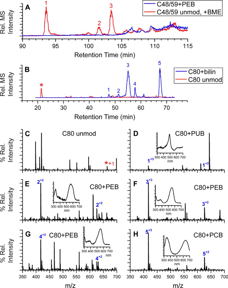
Extracted ion chromatograms and LC-MS spectra for trypsin-digested peptides from ΔcpeF PE. A, combined extracted ion chromatograms for the peptides RLDAVNAIASNASC48MVSDAVAGMIC59ENQGLIQAGGNCYPNR at m/z 1200. 06 ((M + 4H)4+, PEB-modified; blue line) and m/z 1404.32 ((M + 3H)3+, unmodified (unmod) + one BME; red line) of CpeB isolated from ΔcpeF cells grown in green light. Numbers 1–3 indicate three versions of the unmodified peptide observed (one BME on Cys-48, -59, or -71). B, extracted ion chromatograms for the peptides MAAC80LR at m/z 625.81 ((M + 2H)2+, bilin-modified; blue line) and m/z 332.67 ((M + 2H)2+, unmodified; red line and asterisk) of CpeB isolated from ΔcpeF cells grown in green light. Numbers 1–5 indicate the MAAC80LR peptides that have a bilin bound. C, MS of unmodified MAAC80LR. D–H, MS for the five modified versions of the peptide MAAC80LR containing Cys-80 as specified in B. Inset graphs are the UV-visible absorbance spectra for the peaks at the retention times that are numbered with a + charge. The type of bilin bound to Cys-80 is indicated per panel. These results are representative of two independent replicates.

