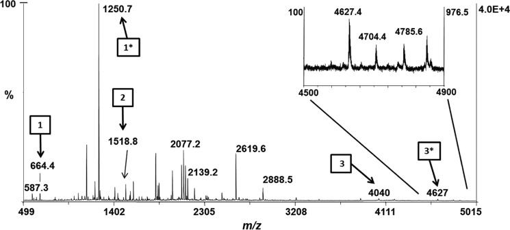
Figure 5.
MALDI-MS spectrum of recombinant HT-CpeB peptides. The MALDI-MS spectrum of the peptide mixture resulting from the trypsin digestion of HT-CpeB purified from E. coli cells expressing pCpeBZ, pCpeS, pHT-CpeF2, and pPebS (BZ+S+F+PEB sample) is shown. Arrows bearing numbers indicate peaks that were identified as tryptic peptides containing a Cys residue without PEB bound: Cys-80 (arrow 1, m/z 664.41+), Cys-165 (arrow 2, m/z 1518.81+), and Cys-48/Cys-59 (arrow 3, m/z 40401+). Arrows bearing numbers with asterisks indicate peaks that were identified as tryptic peptides containing a PEB chromophore. Attachment was found to occur at Cys-80 (arrow 1*, m/z 1250.71+) and double attachment at Cys-48/Cys-59 (arrow 3*, m/z 4627.41+). No attachment to Cys-165 was detected. The inset graph is a close-up view of the m/z 4500–4900 range. These results are representative of two independent replicates.

