Fig. 3.

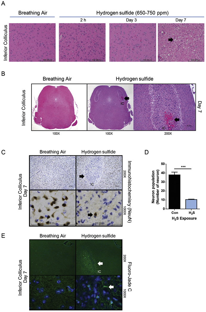
Neurodegeneration and necrosis in the IC of mice exposed to 650–750 ppm H2S in acute short-term repeated inhalation exposures over 7 days. Note the loss of neurons and development of clear vacuoles in the neuropil (arrow) on day 7 in mice exposed to H2S and the minimal morphologic effects to earlier H2S exposures or breathing air (A). H2S exposure induced hemorrhage (thick arrow) in the IC. An insert at higher magnification is included to show the hemorrhage. Hemorrhage was not seen in control mice (B). NeuN staining in brown color (arrow) of neurons (C). H2S caused marked and selective loss of neurons in the IC (1000× magnification images, C), with retention of neurons in the regions surrounding the IC (200× magnification image) (C). Computer aided image analysis of NeuN immunostained sections reveals marked loss of neurons (D) in the IC of mice exposed to H2S (p < 0.001, t-test). Representative photomicrographs of mice exposed to breathing air or H2S, hematoxylin and eosin (A) and NeuN immunohistochemistry (C). Neurons in the IC region of the H2S exposed mice on day 7 were enumerated and compared to breathing air control (D). Degenerating neurons were visualized with Flouro-Jade C staining (E). Arrow indicates degenerative neurons. Neurodegeneration was not seen in the control group. (For interpretation of the references to color in this figure legend, the reader is referred to the web version of this article.)
