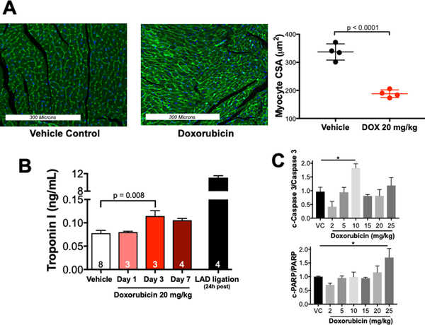
Figure 2. Doxorubicin causes cardiomyocyte atrophy in vivo.
Mice were sacrificed 7 days after receiving doxorubicin 20 mg/kg i.p. (A) Representative wheat germ agglutinin-Alexa Fluor 488 conjugate stained heart sections. Analysis of myocyte cross-sectional areas from 4 hearts (800 cross-sectional areas total, 200 cross sections per mouse over 4–6 histological sections). A Student’s t-test was used to determine significance between groups. (B) Serum Troponin I was measured by ELISA. (C) Summary densitometry of immunoblots for markers of apoptosis. n= 6 for VC, n = 3–4 for DOX-treated. Results were compared using a one-way ANOVA. c-Caspase 3 = cleaved caspase 3; c-PARP = cleaved poly ADP ribose polymerase, LAD = left anterior descending coronary artery.

