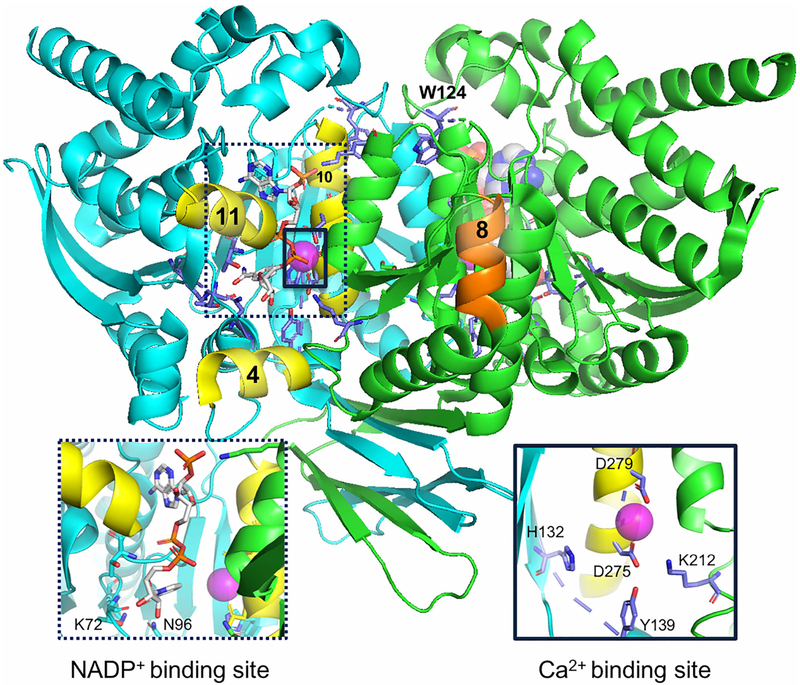
Figure 2. IDH1 dimeric structure (PDB: 4KZO [31]).
α-Helices 4, 8, 10, and 11 are labeled for reference and colored in yellow (monomer A, helices 4, 10, and 11) and orange (monomer B and helix 8). The NADP+- and Ca2+-binding sites are expanded in the labeled sub-panels. Calcium is shown as a pink sphere. NADP+ is colored based on atoms, with nitrogen (blue), oxygen (red), phosphorus (orange), and carbon (gray) shown. Image was generated with PyMol [63].

