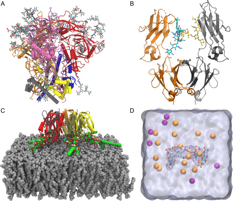
Fig. 4.
Practical applications of Glycan Reader & Modeler. (A) HIV envelope protein trimer of heterodimer with N-glycans. Each heterodimer is composed of gp120 (gray, blue, and yellow) paired with gp41 (orange, red, and pink, respectively). (B) Immunoglobulin G Fc dimer (orange and gray) with N-glycans. (C) Cholera toxin B and ganglioside GM1 pentameric complex shown with each protein segment colored differently. GM1 glycolipids are shown as green sphere. A membrane bilayer (gray) is composed of DMPC. (D) Heparan sulfate pentasaccharide surrounded by K+ (orange) and Cl– ions (purple). Water box is shown as a translucent surface.

