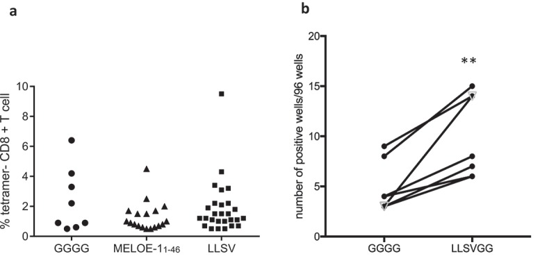
Figure 6.

Panel A. Typical example of an in vitro stimulation of PBMC from a healthy donor with MELOE-1 SLP (MELOE-113-27 xxxx MELOE-136-44) containing various linkers or the native MELOE-111-46 followed by the assessment of CD8 responses (number of positive microcultures and frequency of positive tetramer+/CD8+ lymphocytes per well). PBMC were stimulated in 96 well plates with SLP (5 µM) and cytokines (see M&M) for 21 days. Microcultures were screened by tetramer and CD8 double staining (threshold 0.5% of positive cells). Panel B. Comparison of in vitro stimulation with the same aSLP as in A containing either the linker GGGG or the linker LLSVGG with PBMC from six healthy donors and one melanoma patient (open triangle). **p = 0.004, paired t-test.
