Figure 1.

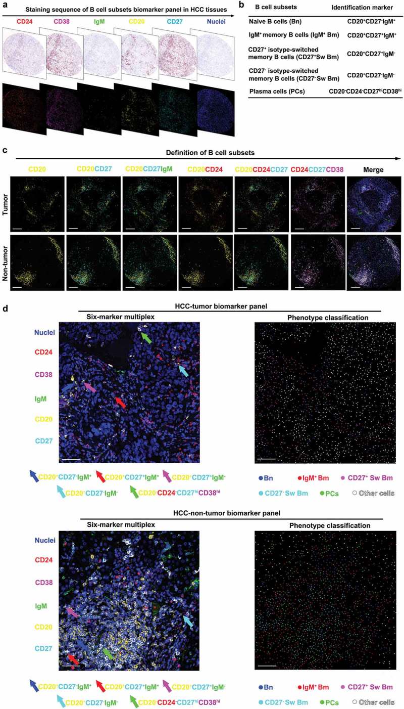
B cell subsets are defined by six-color multiplexed immunohistochemistry in HCC. (a) Digital scanning displayed bright-field image and multispectral image (MSI) of one TMA core from HCC tissues. (b) B cell subsets and corresponding identification markers applied in this study. (c) The multiplexed images displayed co-localization of different markers. Scale bar: 200 μm. (d) The representative images of six-marker multiplex and phenotype classification. Scale bar: 50 μm.
