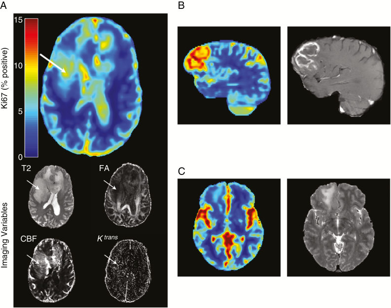
Fig. 3.
Quantitative maps. Ki-67 map generated using the random forest trained on the 4 imaging inputs shown in axial view: T2-weighted image, FA, Ktrans transfer constant, and CBF. Areas of increased estimated Ki-67 (white arrow) likely contain more actively dividing tumor cells and provide a better target for biopsy. In tumors like (A), a grade III anaplastic astrocytoma, sampling the most malignant area of the tumor is important for correctly grading the tumor. This tumor did not demonstrate contrast enhancement, which is clinically used for biopsy target selection, when present. (B) for enhancing tumors (glioblastoma multiforme shown), the highest estimated Ki-67 generally corresponds to enhancement (the currently used clinical target for biopsy selection). (C) For low-grade tumors (shown grade II diffuse glioma) the estimated (and measured) Ki-67 is generally low. Seemingly raised Ki-67 values in the sulci and cisterns are artefactual, but are unlikely to confuse an experienced clinician.

