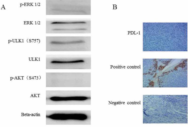
Figure 3.

Molecular analysis of the biopsy tissue from lung metastasis. (A) The phosphorylation of ERK1/2 and AKT(S473)was weak in the metastatic tissue,and the phosphorylation of ULK-1 S757 was relatively strong. (B) The PDL-1 expression was negative in the biopsy tissue of lung metastases (IHC 10 × 10).
