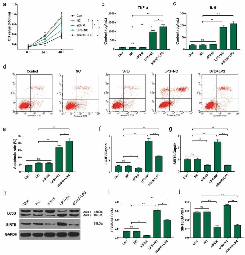
Figure 6.

Effect of SIRT6 silencing on HK-2 cells. (a) The CCK-8 assay was applied to detect cell viability. (b–c) ELISA was used to test the concentration of TNF-α and IL-6 in culture. (d–e) Apoptosis rate was detected using flow cytometry. (f–j) SIRT6 and LC3B mRNAs and proteins were detected by QRT-PCR and Western blot. *P< 0.05,**P< 0.01. (biological replicate = 12, technical replicate = 3).
