Figure 2.

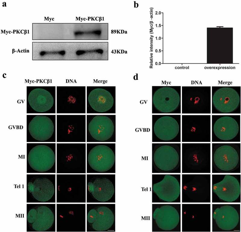
Expression and subcellular localization of Myc-PKCβ1 during mouse oocyte meiotic maturation. (a) Myc-PKCβ1 expression in oocytes injected with Myc-PKCβ1 mRNA. Myc mRNA and Myc-PKCβ1 mRNA injected oocytes were collected for Western blot analysis. After microinjection of PKCβ1-myc mRNA (2.5mg/ml) or Myc mRNA, oocytes were incubated in M2 medium containing 200μm IBMX for 12h, and then collected for Western blot analysis. Each sample contains 200 oocytes. (b) The intensity of Myc-PKCβ1/β-actin was accessed by grey level analysis. The molecular mass of Myc-PKCβ1 was 89 kDa. (c) Myc-PKCβ1 mRNA-injected oocytes at GV, GVBD, MI, TI and MII stages were stained with anti-Myc antibody. Green, Myc-PKCβ1; red, chromatin; each sample was counterstained with Hoechst 33,342 to visualize DNA (red). Bar = 20μm. (d) Confocal microscopy of showing the subcellular localization of Myc (green) in myc mRNA-injected oocytes at GV, GVBD, MI, T1 and MII stages. Green, Myc; red, chromatin. Bar = 20μm.
