Figure 3.

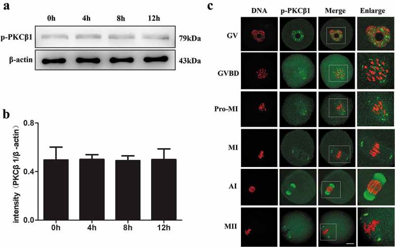
Expression and subcellular location of p-PKCβ1 during oocyte meiotic maturation. (a)Expression of p-PKCβ1 protein identified by Western blotting. Samples of 200 oocytes were collected after culture of 0, 4, 8 and 12 h, corresponding to GV, GVBD, MI and MII stages, respectively. The molecular mass of p-PKCβ1 and β-actin were 79kDa and 43kDa, respectively. Each sample contained 200 oocytes. (b)The intensity of p-PKCβ1/β-actin was accessed by grey level analysis. (c) Confocal microscopy showing the subcellular localization of p-PKCβ1 at GV, GVBD, Pro-MI, MI, AI and MII stages. Green, PKCβ1; red, chromatin; each sample was counterstained with Hoechst 33,342 to visualize DNA (red). Magnifications of the boxed regions are shown on the right of each main panel. Bar = 20μm.
