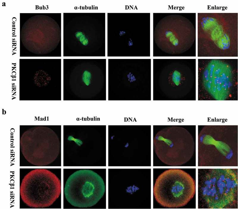
Figure 6.

PKCβ1 depletion caused activation of SAC. Oocytes were arrested in M2 medium containing 200μm IBMX for 24h, following injection of PKCβ1 siRNA or control siRNA, then washed and cultured in IBMX-free M2 medium for10h. (a) Bub3 as marker of SAC was detected at the kinetochores in the PKCβ1-depletion group. Red, Bub3; green, α-tubulin; blue, DNA. Bar = 20μm. (b) Mad1 staining was used to further confirm the phenotype. Red, Mad1; green, α-tubulin; blue, DNA. Bar = 20μm.
