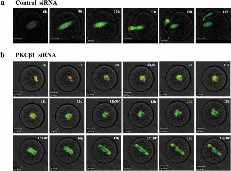
Figure 7.

PKCβ1 knockdown disrupted the metaphase-anaphase transition of mouse oocytes as revealed by time-lapse live cell imaging. (a)Oocytes were co-injected with β5-tubulin-GFP mRNA, H2B-RFP mRNA and control siRNA. Spindle (fluorescent tubulin) and DNA (red) images in a representative control oocyte during in vitro maturation. Time points indicating the time lapse from GVBD occurrence in the oocytes. (b)Similar to (A), oocytes were co-injected with β5-tubulin-GFP mRNA, H2B-RFP mRNA and PKCβ1 siRNA. Representative images showing the PKCβ1 depleted oocytes with abnormal spindles, Pro-MI/MI arrested chromosomes, misaligned chromosomes, repetitive and unsuccessful chromosome segregation and PB1 extrusion failure. Green, tubulin; red, DNA. Bar = 20μm.
