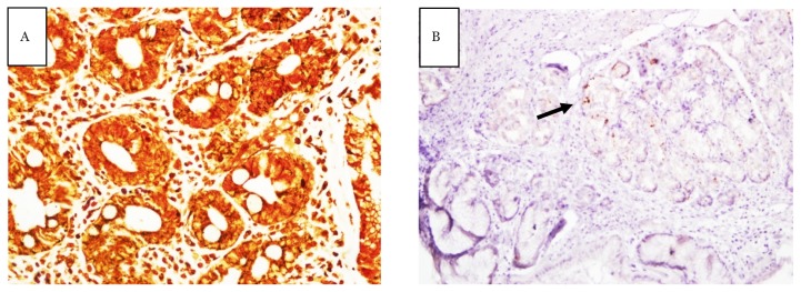
Figure 2.
Slide stained with WS staining at 40× magnification (A) and slide stained with IHC at 10× magnification (B); both were from the same gastric biopsy tissue. Bacteria appeared as fragmented and misreported as negative WS staining. Confirmation with the gold standard staining (IHC) showed positive H. pylori infection (arrow)

