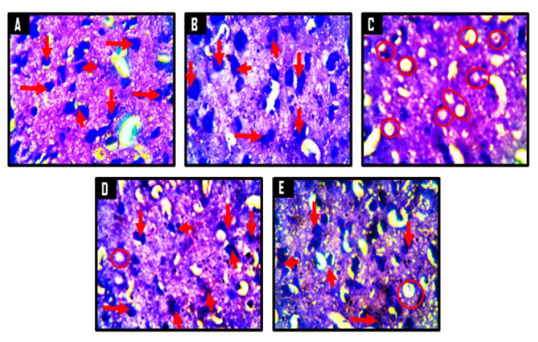
Figure 13.
Representative photomicrographs of the internal molecular layer of the prefrontal cortices of Wistar rats showing the Nissl substances in the neuronal cells. A = control, B = corn oil (CO), C = cuprizone (CPZ), D = cuprizone then kolaviron (CPZ then Kv) and E = kolaviron then cuprizone (Kv then CPZ). A and B showed abundant Nissl substances within the neurons (red arrows), while C showed severe chromatolysis as indicated by poorly stained Nissl substances in the neurons (red circles). D and E presented with mild chromatolysis relative to C. Cresyl Fast Violet Stain ×400

