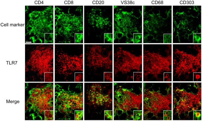
Figure 2.

Co‐localization of Toll‐like receptor (TLR)‐7 and mononuclear cells (MNCs) in LSGs from the pSS patients. Representative samples of LSGs stained with cell markers for T cells (anti‐CD4 and anti‐CD8 antibodies), B cells (anti‐CD20 antibody), plasma cells (anti‐VS38c antibody), macrophages (anti‐CD68 antibody) or pDCs (anti‐CD303 antibody) (green), and anti‐TLR‐7 antibody (red) from pSS patients (n = 3). Hoechst (blue) was used for counterstaining the nuclei. Insets: representative staining for each panel. Bar: 40 μM.
