
FIG. 2.
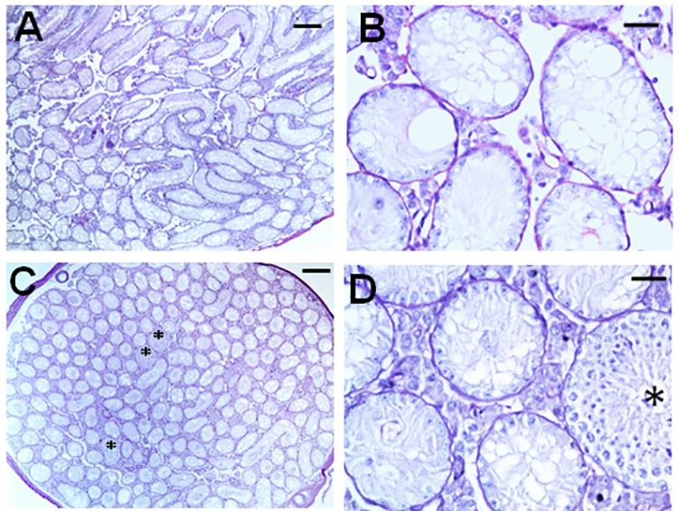
Representative histology of the testis in B6 mice at 4 weeks (A&B) and 8 weeks (C&D) after testicular irradiation with total doses of 13.5 Gy. B and D are the magnified views of areas in A and C, respectively. Asterisks in C &D represent the tubules showing differentiated germ cells derived from a few endogenous radioresistant A spermatogonia. Note that at 4 weeks, the tubules with differentiated germ cells are almost absent. The bars indicate 200 μm in A&C and 30μm in B&D.