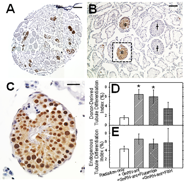
FIG. 3.

Testicular histology (A-C) and analysis of tubules with spermatogenesis (D-E) after syngeneic (B6 GFP mouse to B6 mouse) transplantation. (A) Testicular section 8 weeks after transplantation. Donor colonies were immunolocalized with anti-GFP followed by DAB staining and then counterstained with hematoxylin. (B) Another section at higher magnification with tubules showing endogenous (†) and donor-derived (*) spermatogenesis. (C) Higher magnification of tubule with donor derived spermatogenesis showing the presence of spermatid and sperm. Percentages of tubules with (D) donor-derived and (E) endogenous spermatogenesis after transplantation to recipients with different hormone manipulations. The number of recipient testes analyzed in each group were 10–12. Significant differences in the tubule differentiation indices between different groups and irradiated-only mice by ANOVA and post-hoc Tukey’s test are indicated (* = P<0.05). In addition, the differentiation of donor cells in the group receiving GnRH-ant plus FSH was significantly lower than in the group only receiving GnRH-ant by non-parametric tests. The bars indicate 350 μm in A, 100 μm in B and 25μm in C.
