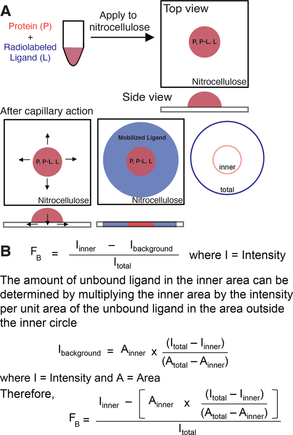
Figure 1.
Principle of DRaCALA.
(A) Schematic representation of DRaCALA assay on application of protein-ligand mixture (in red) onto nitrocellulose with consecutive ligand mobilization (in blue) by capillary action. Protein (P), ligand (L), and protein–ligand complex (P•L) distribution during the assay is shown. (B) Equations used to analyze DRaCALA data for fraction of the ligand bound (FB) for purified proteins. Taken with permission from Roelofs et al. (2011).

