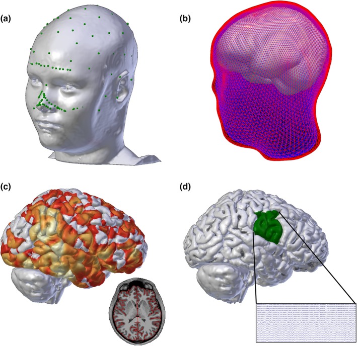
Figure 2.

(a) EEG electrode coordinates (green) are co‐registered to a detailed head model generated by each subject's MRI. (b) Boundary element models of the outer skin (red), outer skull (blue), and inner skull (white) layers are generated for the forward solution model. (c) Example sourcedata at one time point; the solution is constrained to the cortex as shown in the axial MRI slice inset (colors indicate current magnitude). (d) Source data in the seizure onset zone (e.g. the lower half of the sensorimotor cortex) are selected for analysis (example cortical source activity time series recording shown in inset)
