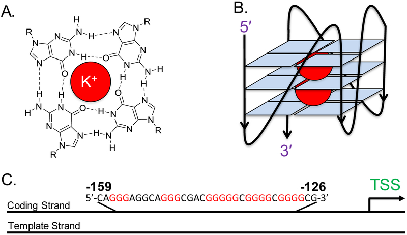
Figure 2.
(A) The structure of a G-tetrad, in which four guanine bases associate via Hoogsteen pairing around a K+ ion, represented by the red circle. (B) A G4 representation, in which three tetrads are stacked on top of one another and stabilized by two K+ ions to adopt a parallel-stranded G4 fold. (C) The PCNA PQS showing its position in the coding strand between −126 and −159 relative to the transcription start site (TSS) of the gene.

