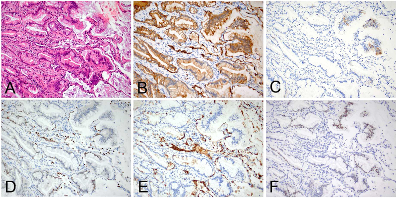
Figure 12.
An example of invasive mucinous adenocarcinoma of the lung demonstrating lepidic and acinar patterns (A), diffuse CK7 expression (B), focal CK20 expression (C), scattered foci with weak TTF1 (D) and/or Napsin-A (E) expressions, and weak to moderate CDX2 expression (F). Of note, the entrapped type II pneumocytes are reactive to CK7 (B), TTF1 (D) and Napsin-A (E).

