Figure 3.

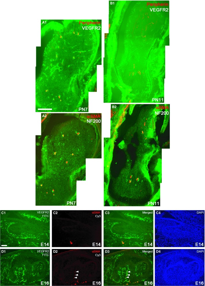
Comparison of the localization of blood vessels and neurites in the mandibular first molar tooth germ at selected postnatal stages (PN7, PN11) detected by immunofluorescence using anti‐VEGFR2 and anti‐peripherin antibodies (A1, B1). Anti‐αSMA and anti‐NF200 antibodies were used to display association of neurites with blood vessels with αSMA‐immunopositive mural cells (A2, B2). Sections are approximately from the level of the future enamel‐cementum junction. (Horizontal sections) Scale bar in (A1): 100 μm; applies to (A1–C2). (D1–E4) Blood vessels and adjoining smooth muscle cells in sagittal sections of mandibular first molar tooth germs at E14 and E16, detected by immunofluorescence using anti‐VEGFR2 (C1, D1) and anti‐αSMA antibodies, respectively (C2,D2). (C4, D4) DAPI staining to display cell nuclei. C4 and D4 show merged channels. Blood vessels with no smooth muscle cells accompanying them are seen in the E14 dental follicle and papilla. The inferior alveolar artery is enveloped by smooth muscle cells (C1–C3). Two blood vessels with aligned smooth muscle cells are observed to invade E16 dental pulp (white arrowheads) (D1–D3). Scale bar in (C1): 100 μm; applies to (C1–D4).
