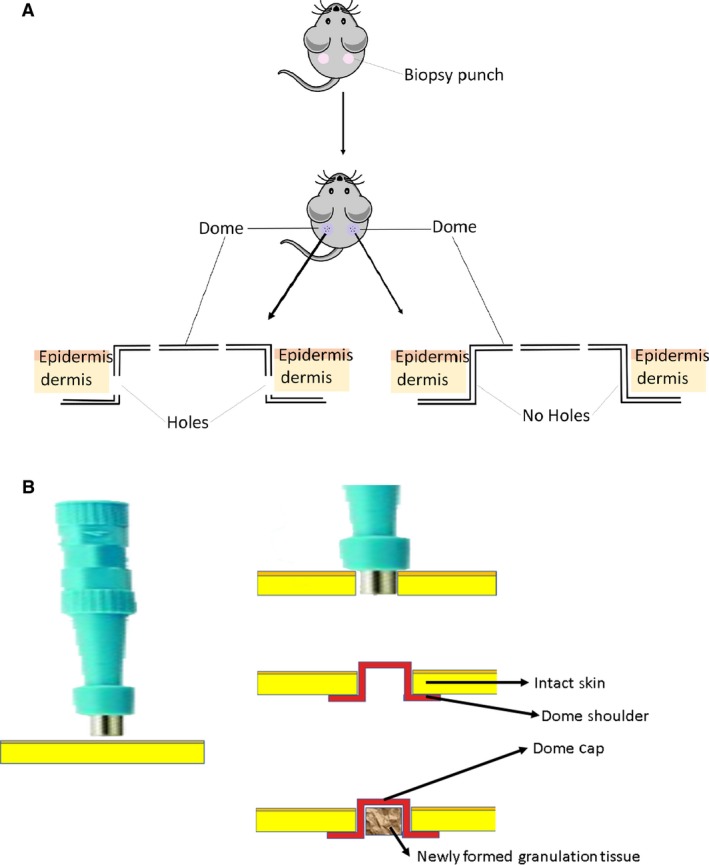
Figure 2.

Infliction of wound and administration of dome. (A) Excisional biopsy punches were inflicted to produce one wound on each side of the mouse of equal diameter size. The nonperforated dome was added to the right‐side wound while the perforated domes were added to the left‐side wound on each mouse. The shoulders of the dome were placed for 11 days under the surrounding skin, preventing contraction. The wounds were harvested for analysis. (B) Insertion of the dome into the skin after punch biopsy and formation of the granulation tissue, which mainly comprises vascular connective tissue.
