
Figure 3.
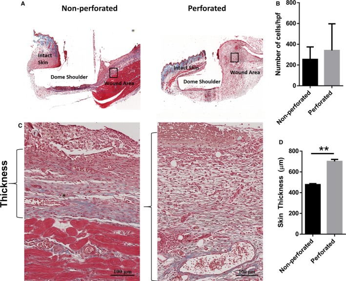
Newly formed granulation tissue is thicker in wounds with a perforated dome. (A) Masson's trichrome staining was performed on cross‐sectional slices of the harvested wounds. Representative images of the formed granulation tissue after 11 days can be seen in (C). Brackets indicate the measured thickness of the skin. (B) No significant differences in cellularity were observed between the non‐perforated and perforated dome groups. (D) Skin thickness was significantly higher in the perforated group compared to the barricaded model (n = 3, P < 0.05, unpaired t‐test).