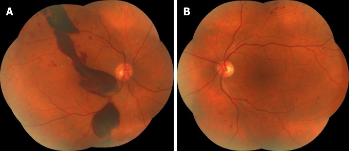
Figure 3.
Colored fundus photographs of a diabetic patient reveal the progression of diabetic retinopathy in the right eye after surgery. A, B: Both eyes have dot-blot hemorrhages and hard exudates; two months after surgery, massive preretinal hemorrhages occurred in the right eye (A). A: Right eye; B: Left eye.

