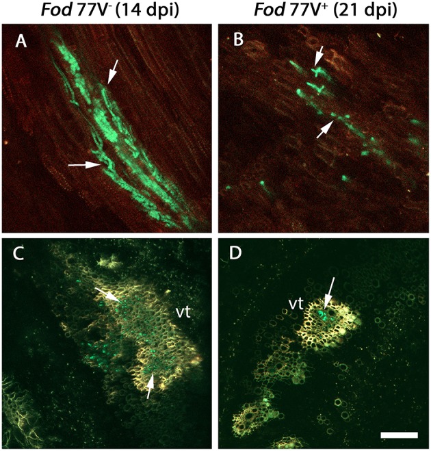
Figure 2.

Colonization of the root crown by the virus-free (V−) and the virus-infected (V+) GFP- strains of F. oxysporum f. sp. dianthi isolate 77. (A) Longitudinal and (C) transversal root crown sections of plants inoculated with the virus-free strain V− (green) 14 days after inoculation. (B) Longitudinal and (D) transversal root crown sections of plants inoculated with the virus-infected strain V+ (green) 21 days after inoculation. Both, the virus free and the virus-infected strain, colonized the plant vascular tissue (arrowed), but the number of vessels colonized as well as the number of hyphae inside them were lower in plants inoculated with the strain carrying the virus. vt, vascular tissue. Scale bar = 50 μm (A,B) and 100 μm (C,D).
