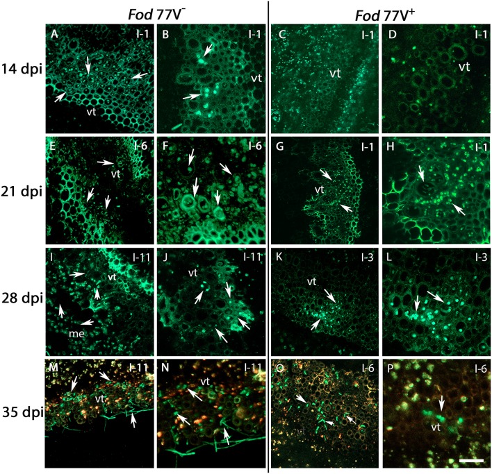
Figure 3.
Internal colonization of stem internodes after inoculation of carnation plants with the virus-free (V−) and the virus-infected (V+) GFP-strains of F. oxysporum f. sp. dianthi isolate 77. Transversal sections of the internodes (I) were obtained at 14, 21, 28, and 35 days post inoculation (dpi), and were analyzed by confocal laser scanner microscopy. (A,B) Hyphae of the virus-free strain were detected in the vascular tissue of the first internode (I-1) at 14 dpi. (C,D) At the same sampling time (14 dpi), hyphae of the virus–infected strain were not detected yet in the same internode. (E,F) Stem sections of the sixth internode (I-6) showing the vascular tissue extensively colonized by hyphae of strain V− at 21 dpi. (G,H) At the same time point, hyphae of strain V+ only reached the first internode (I-1). (I,J) Stem sections of plants inoculated with the strain V− at 28 dpi showing the hyphae reaching the last stem internode (I-11) of the plant. (K,L) At the same sampling time (28 dpi), the strain V+ only reached the vascular tissue of internode 3 (I-3). (M,N) Extensive colonization of the vascular tissue up to the last plant internode (I-11) by the virus free strain at 35 dpi. (O,P) In contrast, colonization by the V+ strain did only reach internode 6 (I-6) at the same sampling time (35 dpi). Arrows indicate the presence of hyphae (green). vt, vascular tissue. Scale bar = 50 μm (A,C,E,G,I,K,M,O), 20 μm (B,D,F,H,J,L,N,P).

