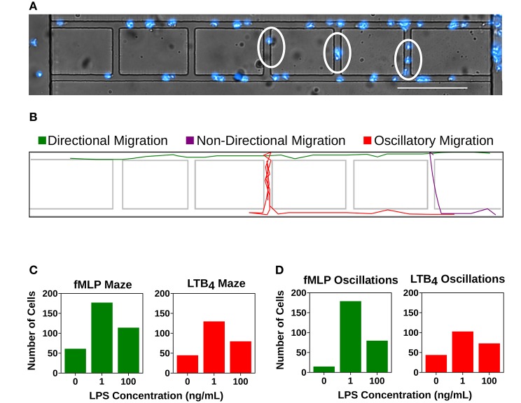
Figure 5.
Super-low dose LPS primes neutrophils for dysfunctional chemotaxis. Quantification of cells entering the mazes within the microfluidic device or displaying an oscillatory migration pattern. (A) dHL-60 cells (blue) migrating within the microfluidic device. Cells within the linear migration channel are migrating directionally toward the chemoattractant gradient. Circled cells are displaying non-directional migration as they are migrating within the mazes. Scale Bar = 150 μm. (B) Computational recreation of cell migration tracks depicting directional migration (green), non-directional migration (purple), and oscillatory migration (red). (C) Number of “lost” cells that entered the maze while migrating toward fMLP (green) and LTB4 (red) in the unstimulated group (fMLP: n = 61, LTB4: 44), the group stimulated with super-low dose LPS (fMLP: n = 177, LTB4 = 129), and the group stimulated with high-dose LPS (fMLP: n = 114, LTB4: 79). Super- low dose LPS priming significantly increases the number of “lost” cells in both fMLP and LTB4 conditions compared to both control and high dose LPS treatment groups. (D) Number of cells that displayed oscillatory migration patterns while migrating toward fMLP (green) and LTB4 (red) in the unstimulated group (fMLP: n = 14, LTB4: 43), the group stimulated with super-low dose LPS (fMLP: n = 178, LTB4 = 102), and the group stimulated with high-dose LPS (fMLP: n = 79, LTB4: 72). Super-low dose LPS priming significantly increases the number of oscillatory cells in both fMLP and LTB4 conditions compared to both control and high dose LPS treatment groups. Data is representative of one experiment, however experiment was repeated at least 3 times.

