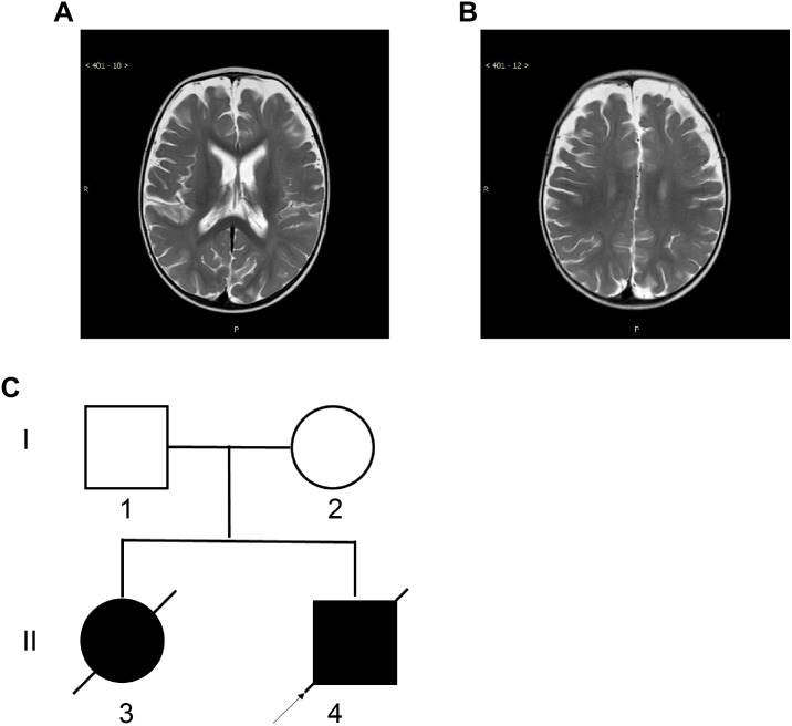
FIGURE 1.
Magnetic Resonance Imaging (MRI) of the proband and the two-generation pedigree. (A,B) Brain MRI of the proband at the age of 10 months showed deep sulci in the frontal and parietal lobes and a wide subarachnoid space. (C) The two-generation pedigree of the family with CMS-EA. The parents are unaffected, while the two offspring are affected. The arrow indicates the proband.

