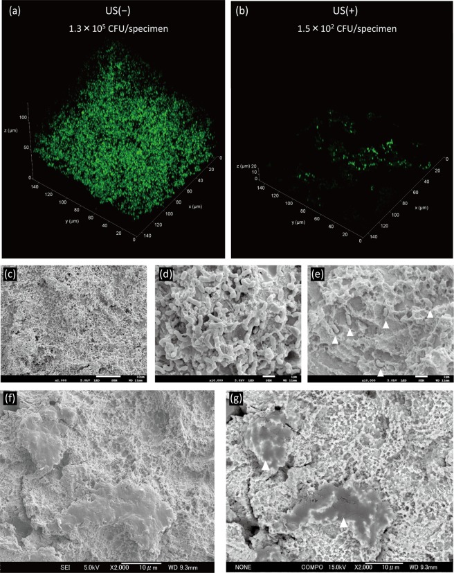
Figure 3.
Representative confocal laser scanning microscopy and scanning electron microscopy images of A. actinomycetemcomitans biofilms formed on titanium specimens treated with or without ultrasound scaling (US). (a) A. actinomycetemcomitans biofilm formed on titanium specimens, and (b) US treatment of the biofilm at a field view of 148 × 148 µm. (c) Biofilm at low magnification. Scale bar = 10 µm. (d) Biofilm at high magnification. Scale bar = 1 µm. (e) Remaining bacteria after US. Scale bar = 1 µm. White arrowheads indicate bacterial cells. (f) Secondary electron image of a titanium surface after US. Scale bar = 10 µm. (g) Backscattered electron image of (f). Scale bar = 10 µm. White arrowheads indicate remnants of the plastic scaler tip.

