Figure 5.
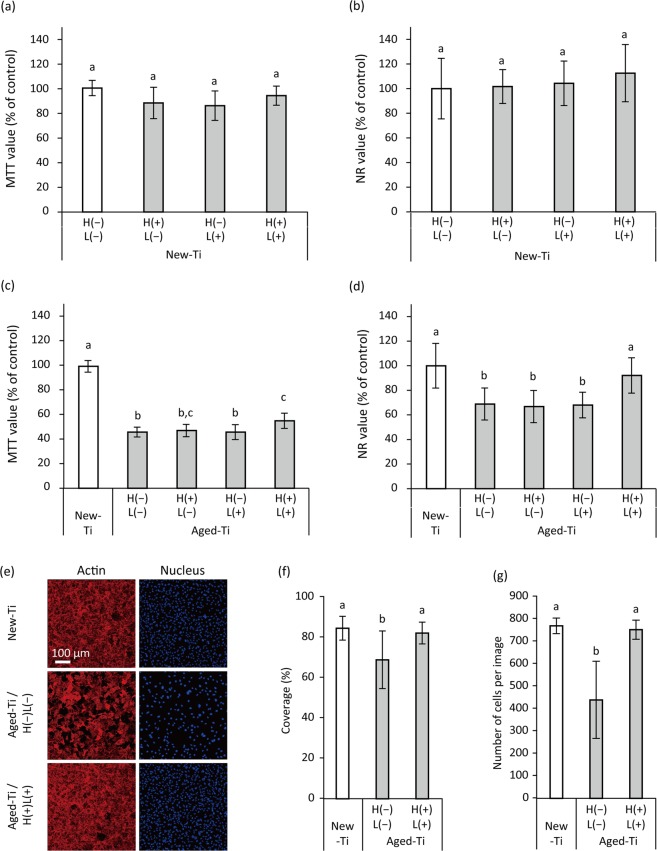
Proliferation of MC3T3-E1 osteoblastic cells cultured on H2O2 photolysis-treated newly prepared (New-Ti) and aged titanium (Aged-Ti), as assessed by methyl thiazolyl tetrazolium (MTT) assays, neutral red (NR) assays, and confocal laser scanning microscopy (CLSM). Cells were cultured on the specimens for 3 d before assays were performed. (a,b) Relative optical density values obtained in MTT assay (MTT value) and NR assay (NR values) for cells grown on New-Ti and (c,d) Aged-Ti. The titanium discs were immersed in 3% H2O2 and irradiated with 365 nm LED, either alone or in combination denoted as H(−)L(−), H(+)L(−), H(−)L(+), or H(+)L(+), for 5 min. (e) Representative CLSM images of MC3T3-E1 osteoblastic cells cultured on New- Ti and Aged-Ti treated with H(−)L(−) and H(+)L(+). Cell nuclei and actin filaments were stained with DAPI and rhodamine phalloidin, respectively. (f) Quantitative results of cellular coverage and (g) number of cells. Parameter values for cell proliferation on Aged-Ti were significantly lower than those for cells grown on New-Ti. However, H(+)L(+) treatment recovered the reduction in cell proliferation induced by aging. Values and error bars in the graphs indicate the mean and standard deviation, respectively [n = 6 for (a–d) and n = 9 for (f,g)]. Different letters above the columns in the graphs refer to significant differences (p < 0.05) between different groups. H(−)L(−), treatment with pure water in a light-shielding box; H(+)L(−), treatment with 3% H2O2 in a light-shielding box; H(−)L(+), 365-nm LED irradiation of sample in pure water; H(+)L(+), 365-nm LED irradiation of sample in 3% H2O2.

