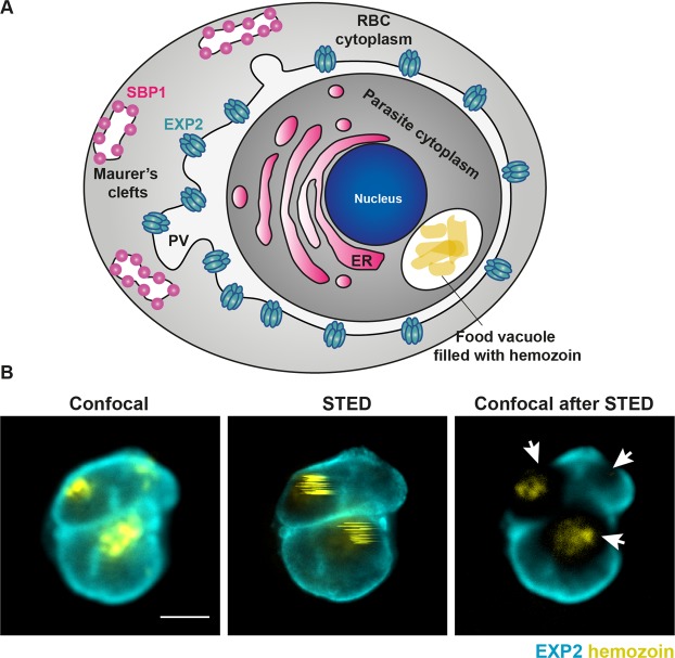
Figure 1.
The hemozoin challenge in malaria samples. (A) Schematic representation of a red blood cell infected with human malaria parasite Plasmodium falciparum at a mid- trophozoite stage. The position of hemozoin-rich food vacuole has been marked as well as several parasite-derived structures important for parasite protein export into the host cell. ER – endoplasmic reticulum, PV – parasitophorous vacuole surrounding the parasite, RBC – red blood cell, EXP2 – the core component of parasite protein translocon, SBP1 – a marker of Maurer’s clefts which are parasite-derived structures that facilitate protein trafficking to the surface of infected red blood cell. (B) Confocal and conventional STED images image of an infected red blood cell stained for EXP2 marking parasitophorous vacuole (cyan) and hemozoin in the food vacuole visualised via reflected light of the STED laser (yellow). Conventional STED increases the resolution but damages hemozoin and any surrounding cellular structures as evidenced in confocal images recorded after a single frame of STED imaging. Scale bar 2 μm.

