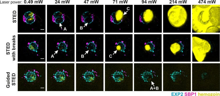
Figure 3.
Sample damage in conventional or guided STED. Representative confocal images of infected red blood cells stained for the parasitophorous vacuole (EXP2, cyan) and Maurer’s clefts (SBP1, magenta) and hemozoin visualisation (yellow) using reflected light from the excitation laser. Images were recorded after single rounds of STED imaging with increasing powers (0.49, 24, 47, 71, 91, 214 or 474 mW) of the STED laser to compare damage inflicted in conventional STED mode, conventional STED mode with illumination breaks and guided STED mode. Examples of three major stages of sample damage: A – altered hemozoin structure, B – damages of fluorophores in the proximity of hemozoin, C – hemozoin disintegration. Scale bar 2 μm.

