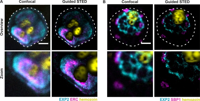
Figure 4.
Two-colour imaging of parasite structures using guided STED. Confocal and guided STED images of infected red blood cells. Staining for EXP2 (cyan) and ERC (magenta, A) or for EXP2 (cyan) and SBP1 (magenta, B). Lower panels show a magnified view of respective structures at super-resolution. The ER (magenta, A) and parasitophorous vacuole (cyan, A and B) are located close to the hemozoin particles (yellow) which are visualised via reflected light of the STED laser. Guided STED prevented sample damage by switching off the STED laser in areas with high amounts of hemozoin. Scale bar 2 μm.

