Figure 1.
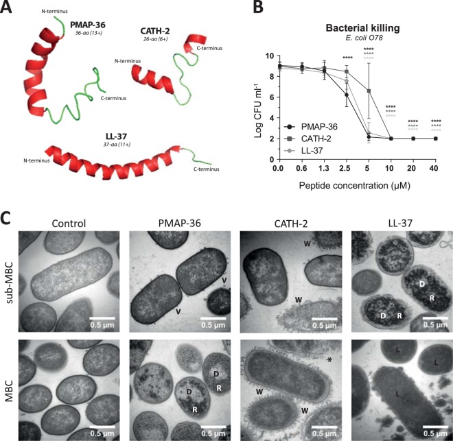
PMAP-36, CATH-2 and LL-37 efficiently kill E. coli O78 but in a different way. Structures of PMAP-36, CATH-2 and LL-37 as predicted using I-TASSER46–48 (A). Bacterial killing of 106 CFU ml−1 E. coli O78 by PMAP-36, CATH-2 and LL-37 was tested using a colony count assay (B). Peptide dependent morphological changes of E. coli O78 were determined by TEM. Representative images are shown for the MBC value and 4x below MBC value (sub-MBC). V – vesicle; D – clustered DNA; R – clustered ribosomes; W – detached and wrinkled membrane; * - ruptured cell; L – lysed cell (C). Data is plotted as average +/− s.d. (N = 3–4). Samples were compared to the no peptide control, using two-way ANOVA with the Bonferroni post-hoc test. (*p ≤ 0.05; **p ≤ 0.01; ***p ≤ 0.001; ****p ≤ 0.0001; black - PMAP-36; dark gray – CATH-2; light gray – LL-37).

