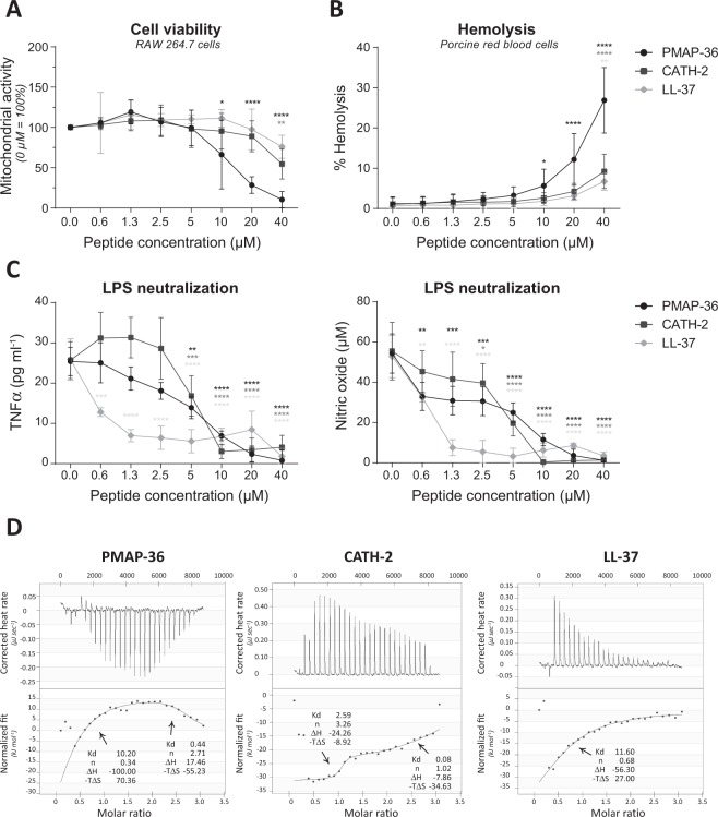
Figure 2.
PMAP-36, CATH-2 and LL-37 exhibit different LPS binding characteristics. Cytotoxic effects against RAW264.7 cells were tested using a WST-1 assay, indicating cell viability. No peptide control was set to 100% cell viability (A). Hemolytic effects on porcine red blood cells were tested by determining heme release. Triton (0.2% v/v) was set to 100% lysis and no peptide as negative control (B). Peptides were mixed with 100 ng ml−1 LPS O111:B4 for 5–10 minutes, before cells were stimulated with this mixture. TNFα and NO production were used to measure cell activity (C). Thermodynamic binding capacity of the peptides was measured with isothermal titration calorimetry (ITC). Every 300 seconds, 1.96 μl peptide solution (200 μM) was titrated into 164 μl LPS solution (25 mM). Heat evolved was measured (top panel) and normalized integrated heat was plotted against the molar ratio between LPS and the peptides (lower panel). Two independent models were used to fit the data and to calculate Kd (μM), the amount of peptide that binds to LPS (n), ΔH (kJ mol−1) and −TΔS (kJ mol−1). Experiments (N = 2) were corrected for heat change of dilution (buffer into buffer titration) and averaged before plotting and model fitting (D). Data is plotted as average +/− s.d. with N = 5–7 (A,B) or N = 3 (C). Samples were compared to the no peptide control, using two-way ANOVA with the Bonferroni post-hoc test. (*p ≤ 0.05; **p ≤ 0.01; ***p ≤ 0.001; ****p ≤ 0.0001; black - PMAP-36; dark gray – CATH-2; light gray – LL-37).

